Keratoconus Treatment Specialist
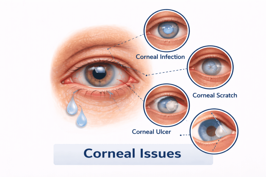
Keratoconus is an eye condition where the normally round, dome-shaped cornea (the clear front surface of the eye) gradually thins and bulges outward into a cone shape. This irregular shape can distort vision and make everyday tasks like reading or driving more difficult.
Causes
The exact cause isn’t fully understood, but several factors may play a role:
- Genetics (family history of keratoconus)
- Chronic eye rubbing
- Underlying conditions like allergies, asthma, Down syndrome, or connective tissue disorders
- Oxidative stress that weakens the corneal tissue
Symptoms
- Blurred or distorted vision
- Increased sensitivity to light and glare
- Frequent changes in glasses prescription
- Difficulty seeing at night
- Eye strain or headaches
- Often affects both eyes, but one eye may be worse
Symptoms usually begin in the late teens to early 20s and may progress over 10–20 years.
Diagnosis
- Comprehensive eye exam
- Corneal topography: A special scan that maps the shape of the cornea (most accurate way to diagnose keratoconus)
- Pachymetry: Measures corneal thickness
- Slit-lamp exam to check for corneal thinning or scarring
Management
Early/Mild Cases:
- Eyeglasses or soft contact lenses to correct vision
Moderate Cases:
- Rigid gas permeable (RGP) or scleral contact lenses to improve focus by creating a smooth surface
- Hybrid lenses (hard center with soft edges) for comfort and clarity
To Slow or Stop Progression:
- Corneal Collagen Cross-Linking (CXL) - A minimally invasive procedure that strengthens the cornea using vitamin B2 (riboflavin) drops and UV light. This can halt progression but doesn't reverse damage.
Advanced Cases:
- Corneal transplant (keratoplasty) may be needed if scarring or thinning becomes severe and vision cannot be corrected with lenses
Prevention Tips
- Avoid eye rubbing—it can make keratoconus worse
FAQ
Keratoconus is a progressive, non-inflammatory eye disease where the clear, dome-shaped outer window of the eye (the cornea) gradually thins and bulges outward into an irregular cone shape.
Common early symptoms include:
- Frequent changes in eyeglass prescriptions, particularly increasing cylindrical (astigmatic) power.
- Distorted or blurred vision where straight lines appear bent.
- Increased sensitivity to bright light and glare (photophobia).
- Halos, starbursts, or ghost images around lights during nighttime driving.
- Eye irritation or headaches linked to chronic eye strain.
No, keratoconus does not cause total black-out blindness because it only alters the cornea and does not damage the optic nerve or retina. However, if left untreated, the progressive thinning and cone bulging can cause severe irregular astigmatism and apical corneal scarring, resulting in significant visual impairment (often classified as "legal blindness") that cannot be corrected with regular spectacles.
The exact cause is multifactorial, involving genetic predisposition, environmental triggers, and cellular enzyme imbalances that weaken corneal collagen fibers:
- Chronic, Vigorous Eye Rubbing: The single largest mechanical trigger. Eye rubbing breaks down collagen cross-links, accelerating corneal thinning and cone formation.
- Ocular Allergies & Atopy: Conditions like allergic conjunctivitis or asthma that prompt intense itching.
- Genetic Factors: Roughly 8% to 10% of patients have a direct blood relative with the condition.
- Connective Tissue Disorders: Conditions such as Ehlers-Danlos or Marfan syndrome.
No. LASIK is strictly contraindicated for anyone with keratoconus or even subclinical/forme fruste keratoconus. LASIK involves slicing a corneal flap and removing underlying tissue, which further weakens an already structurally compromised cornea and can trigger severe, rapid corneal ectasia (uncontrolled bulging). Patients with keratoconus require stabilizing treatments like C3R (cross-linking) or specialty rigid/scleral lenses rather than laser vision correction.
Corneal Collagen Cross-Linking (C3R / CXL) is a minimally invasive procedure designed to halt the progression of keratoconus. During the treatment, riboflavin (Vitamin B2) drops are applied to the cornea and activated with calibrated UV-A light. This photo-chemical reaction creates new chemical bonds (cross-links) between adjacent collagen fibrils, stiffening and strengthening the corneal structure to prevent further cone bulging.
C3R is primarily a stabilizing procedure, not a refractive cure. Its primary goal is to arrest disease progression and prevent the need for a future corneal transplant. While many patients experience mild flattening of the cone and slight visual improvement, most patients still require optical rehabilitation after C3R—such as specialty scleral contact lenses or custom rigid gas-permeable (RGP) lenses—to achieve crisp, sharp vision.
Scleral lenses are large-diameter, rigid gas-permeable lenses designed specifically for irregular corneas. Unlike regular contact lenses that rest directly on the sensitive cornea, scleral lenses:
- Vault over the entire irregular cornea without touching the apex, resting gently on the less-sensitive white part of the eye (the sclera).
- Create a fluid-filled reservoir of sterile saline between the lens and cornea, continuously hydrating the eye and neutralising surface irregularities.
- Provide crisp, distortion-free vision and superior day-long comfort compared to traditional small RGP lenses.
Intacs are microscopic, clear, crescent-shaped polymer inserts surgically placed within the deep middle layers (stroma) of the cornea. By acting like structural architectural supports, they flatten the central cone, reshape irregular curvature, and reduce refractive error. Intacs are often combined with C3R cross-linking to both reshape and biomechanically stabilize the cornea simultaneously.
Corneal transplant surgery is considered an advanced-stage option (required in fewer than 10–15% of cases) when:
- The cornea has become too thin for safe C3R cross-linking (corneal pachymetry below safe thresholds).
- Dense apical scarring has developed in the visual axis due to advanced bulging or healed corneal hydrops.
- The patient has severe contact lens intolerance and cannot achieve functional vision with scleral or RGP lenses.
Deep Anterior Lamellar Keratoplasty (DALK) is the preferred partial-thickness transplant technique because it preserves the patient's healthy inner endothelial layer, virtually eliminating the risk of endothelial graft rejection.
Routine eye exams and standard refraction cannot detect early-stage keratoconus. At ARC Eye Centre, diagnosis is established using high-precision diagnostics:
- Pentacam / 3D Corneal Tomography: High-resolution Scheimpflug imaging that generates 3D elevation maps of both the front (anterior) and back (posterior) corneal surfaces, capturing subclinical cone changes years before vision drops.
- Corneal Pachymetry Mapping: Measures precise thickness across the central, paracentral, and peripheral cornea.
- Corneal Biomechanics & Epithelial Thickness Mapping: Analyzes corneal elasticity and structural resistance.
Our Services
Working Hours
Monday:
9am – 7pm
Tuesday:
9am – 7pm
Wednesday:
9am – 7pm
Thursday:
9am – 7pm
Friday:
9am – 7pm
Saturday:
9am – 7pm
Sunday:
11am – 2pm
( Appointment Only )
How can we help you?
ARC Eye and Retina Centre delivers advanced eye and retina care with expert doctors, modern technology, and compassionate patient-focused treatment.