
Retinal Detachment Treatment
What Is Retinal Detachment?
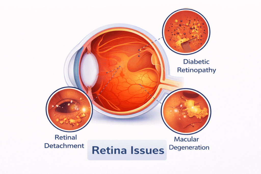
Retinal detachment is a serious eye condition in which the retina, the thin, light-sensitive layer at the back of the eye, separates from its normal position. When this happens, the retina cannot function properly, and vision can be affected. Retinal detachment is an eye emergency, and timely treatment is important to reduce the risk of permanent vision loss.
A retinal detachment may develop when a tear or hole in the retina allows fluid to collect underneath it. In some cases, the retina can be pulled away by scar tissue or other changes inside the eye. It may occur suddenly and without pain.
Some of the early warning signs include flashes of light, a sudden increase in floaters, blurred or reduced vision, or a dark curtain or shadow appearing in the field of vision. These symptoms should not be ignored and require an urgent examination by a Retinal Detachment Specialist.
Diagnosis usually involves a detailed dilated retinal examination, with OCT or ocular ultrasound used when needed. Retinal Detachment Treatment depends on the type, location and extent of the detachment and may include laser treatment, cryotherapy, pneumatic retinopexy, scleral buckle or vitrectomy surgery.
At ARC Eye & Retina Centre in Gurgaon, our retina specialists provide timely diagnosis and advanced Retinal Detachment Treatment, including vitreoretinal surgery, with the aim of preserving and restoring vision wherever possible.
Understanding Retinal Detachment: Signs & Symptoms
Retinal detachment can develop suddenly, and recognising the early warning signs can make a significant difference to vision. The most common symptoms include a sudden increase in floaters, flashes of light or a shadow or dark curtain appearing in the field of vision. Some people may also notice blurred or reduced vision, difficulty seeing in one area or a change in peripheral vision.
A retinal detachment is usually painless, so the absence of pain does not mean the condition is harmless. Symptoms can sometimes begin with a retinal tear before the retina actually detaches.
If you experience new flashes, a sudden shower of floaters or a curtain-like shadow over your vision, you should have your retina examined urgently. Not every floater or flash means retinal detachment, but it is important to rule out a retinal tear or detachment early.
At ARC Eye & Retina Centre, Gurgaon, our retina specialists provide urgent retinal evaluation and timely Retinal Detachment Treatment when required.
Diagnosis and Tests
Diagnosing retinal detachment requires a careful examination of the retina. Your Retinal Detachment Specialist will usually begin with a dilated eye examination to look for retinal tears, holes or areas where the retina has separated from its normal position.
A detailed examination of the peripheral retina is particularly important, as a retinal tear may sometimes be present even when the symptoms are limited. OCT (Optical Coherence Tomography) may be used to assess the macula and understand how much of the retina is affected.
When the retina cannot be seen clearly, such as in the presence of a dense cataract or vitreous haemorrhage, an ocular ultrasound (B-scan) can help determine whether the retina is detached.
The findings from these examinations help the retina specialist decide whether monitoring, laser treatment or surgery is required.
At ARC Eye & Retina Centre in Gurgaon, we use detailed retinal examination and appropriate imaging to diagnose retinal detachment accurately and plan timely Retinal Detachment Treatment.
Why Immediate Retinal Detachment Treatment is Crucial
A retinal detachment should be treated as an eye emergency. Once the retina separates from its normal position, it is no longer receiving the support and blood supply it needs to function properly. The longer the retina remains detached, particularly when the macula is involved, the greater the risk of permanent vision loss.
Early treatment can sometimes prevent a retinal tear from progressing to a complete retinal detachment. If a detachment has already occurred, timely Retinal Detachment Treatment can help reattach the retina and preserve as much vision as possible.
The urgency depends on the location and extent of the detachment, whether the macula is involved and the presence of retinal tears or other complications. This is why sudden flashes, new floaters or a curtain-like shadow should never be ignored.
At ARC Eye & Retina Centre, Gurgaon, we provide prompt retinal assessment and, when required, emergency vitreoretinal care. Our Retinal Detachment Specialists assess each case carefully and recommend the most appropriate treatment without unnecessary delay.
Advanced Retinal Detachment Treatment Options in Gurgaon
The right treatment for retinal detachment depends on the type, size and location of the detachment, the presence of retinal tears and whether the macula is involved. Not every case requires the same procedure.
Small retinal tears or early changes may sometimes be treated with laser photocoagulation or cryotherapy to create a barrier around the affected area and prevent further retinal separation.
For an established retinal detachment, surgical options may include pneumatic retinopexy, scleral buckle surgery or vitrectomy. Vitrectomy is commonly used for more complex detachments and may involve removing the vitreous gel, treating retinal tears and using a gas bubble or silicone oil to help keep the retina in position while it heals.
Your Retinal Detachment Specialist will recommend the procedure based on the individual retinal findings rather than using a single approach for every patient.
At ARC Eye & Retina Centre, Gurgaon, we offer comprehensive Retinal Detachment Treatment and vitreoretinal surgical care using modern retinal imaging and surgical techniques.
Meet Our Expert Retinal Detachment Specialists
At ARC Eye & Retina Centre, Gurgaon, retinal detachment is managed by experienced retina surgeons with specialized training in vitreoretinal care.
Dr. Kritika Singh is a retina and cataract surgeon with postgraduate training from Armed Forces Medical College (AFMC), Pune, followed by advanced fellowship training in vitreoretinal surgery at Aravind Eye Hospital, Madurai. Her practice has a strong focus on retinal diseases and vitreoretinal surgery, along with cataract and comprehensive ophthalmology. Her training and experience allow her to manage a wide range of retinal conditions, including complex retinal detachments.
Dr. Suneel Kumar completed his postgraduate training at the prestigious All India Institute of Medical Sciences (AIIMS), New Delhi. He has extensive experience in the diagnosis and management of retinal and vitreoretinal conditions, with particular expertise in retina, uvea and retinal surgery. His clinical approach focuses on understanding each patient's condition carefully and choosing treatment that is appropriate for their individual needs.
Together, our Retinal Detachment Specialists in Gurgaon provide comprehensive retinal evaluation, Retinal Detachment Treatment and vitreoretinal surgical care, with an emphasis on timely treatment and individualized patient care.
Our Approach to Retinal Detachment Surgery & Recovery
Retinal detachment surgery is planned according to the type and extent of the detachment and the condition of the retina. Before surgery, our retinal team explains the findings, recommended procedure and what you can expect during recovery.
Depending on the case, surgery may involve vitrectomy, scleral buckle or pneumatic retinopexy. During vitrectomy, the vitreous gel may be removed and retinal tears treated with laser or cryotherapy. A gas bubble or silicone oil may be used to support the retina while it heals.
Recovery varies from person to person. Vision may remain blurred for some time after surgery and gradually improve as the retina settles. If a gas bubble is used, certain precautions may be necessary, including avoiding air travel until the bubble has resolved. Follow-up visits are important to monitor retinal healing and eye pressure.
At ARC Eye & Retina Centre, Gurgaon, we provide structured postoperative care following Retinal Detachment Treatment, with regular retinal examinations and guidance throughout recovery to help achieve the best possible outcome.
FAQ
Retinal detachment occurs when the thin, light-sensitive layer of nerve tissue at the back of the eye (the retina) pulls away or separates from its underlying support tissue and blood supply. When detached, retinal cells are deprived of oxygen and essential nutrients. It is considered a sight-threatening ophthalmic emergency because prompt reattachment is critical to prevent irreversible, permanent vision loss.
Retinal detachment itself is painless. The most common warning signs include:
- Flashes of Light (Photopsia): Sudden, bright flickers or sparks, especially in peripheral vision.
- Sudden Shower of Floaters: A rapid increase in dark spots, cobweb-like strands, or specks drifting across your visual field.
- Dark Curtain or Shadow: A fixed, dark veil or shadow beginning in the peripheral vision and moving toward the center.
- Sudden Blurring or Distortion: Visual fuzziness or straight lines appearing wavy.
No. A detached retina cannot heal or reattach on its own. While peripheral retinal tears without detachment can sometimes be sealed using non-surgical laser barrier therapy or cryopexy, once subretinal fluid collects and the retina detaches, surgical intervention is necessary to reattach the retina and preserve vision.
The most frequent causes and predispositions include:
- High Myopia (Severe Nearsightedness): Longer eyeballs stretch and thin the peripheral retina.
- Lattice Degeneration & Retinal Breaks: Thinning patches or untreated holes/tears in the outer retina.
- Posterior Vitreous Detachment (PVD): Age-related shrinking of the vitreous gel tugging on the retina.
- Eye Trauma or Injury: Blunt impact or penetrative ocular trauma.
- Complications of Eye Surgery or Diabetes: Advanced proliferative diabetic retinopathy (tractional detachment) or previous ocular surgeries.
- Family History: Prior detachment in the other eye or hereditary retinal conditions.
Arc Eye Centre provides modern surgical and non-surgical procedures tailored to the detachment's severity:
- Minimally Invasive Vitrectomy Surgery (MIVS): Advanced sutureless, micro-incision vitrectomy (using 23G/25G/27G instruments) to remove the vitreous gel, relieve traction, flatten the retina, and secure it using intraocular gas or silicone oil tamponade.
- Retinal Green Laser Photocoagulation & Cryopexy: Therapeutic laser or freezing probes to seal localized retinal tears and prevent full detachment.
- Scleral Buckling: Placement of a flexible silicone band around the outer eye wall to relieve vitreous traction and reattach the retina.
- Pneumatic Retinopexy: In-office injection of an expandable gas bubble combined with laser/cryo to flatten select superior retinal detachments.
- Vitrectomy: An internal procedure where the vitreous gel is suctioned out from inside the eye to release internal pulling forces, and a gas or silicone oil bubble is placed inside the eye cavity.
- Scleral Buckling: An external procedure where a silicone band is stitched onto the sclera (white part of the eye) from the outside to gently indent the eye wall inward against the detached retina. Depending on the case, a retina specialist may perform one or combine both approaches.
A standard vitreoretinal surgery typically takes 45 to 90 minutes, depending on case complexity and whether silicone oil or gas tamponade is used. The procedure is performed under local or general anesthesia along with intravenous sedation, ensuring the patient feels minimal discomfort during the surgery. Post-operative soreness is manageable with prescribed pain relievers.
When an intraocular gas bubble or silicone oil is placed inside the eye, it naturally floats upward. Maintaining a specific head posture—such as face-down (prone) or side-lying—ensures that the bubble presses directly against the retinal tear, keeping it flat while laser or cryopexy scars form a permanent seal. This positioning is usually maintained for 3 to 10 days as advised by the surgeon.
Visual recovery depends on two key factors:
- Macula Status: If the macula (the center of vision) was still attached before surgery ("Macula-On"), visual prognosis is high. If the macula was detached ("Macula-Off"), central vision may take several months to improve and might not fully return to baseline.
- Time to Treatment: Prompt intervention within 24 to 72 hours of initial symptoms yields the best visual outcomes.
While modern surgical success rates exceed 90%, redetachment can occur in 5% to 10% of cases, most commonly due to Proliferative Vitreoretinopathy (PVR) (scar tissue formation on the retinal surface) or new peripheral tears. If redetachment occurs, an experienced vitreoretinal specialist can perform a revision surgery to re-secure the retina.
Our Services
Working Hours
Monday:
9am – 7pm
Tuesday:
9am – 7pm
Wednesday:
9am – 7pm
Thursday:
9am – 7pm
Friday:
9am – 7pm
Saturday:
9am – 7pm
Sunday:
11am – 2pm
( Appointment Only )
How can we help you?
ARC Eye and Retina Centre delivers advanced eye and retina care with expert doctors, modern technology, and compassionate patient-focused treatment.