
Lattice Degeneration Treatment & Retinal Tears
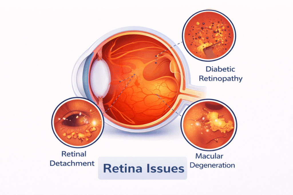
Lattice degeneration and retinal tears are two eye conditions related to changes in the retina, the light-sensitive layer at the back of the eye. They are distinct conditions, both of which can potentially lead to retinal detachment if not properly managed.
1. Lattice Degeneration:
Lattice degeneration is a condition where the retina becomes thinned and weakened, often appearing in a lattice-like pattern. It commonly affects the peripheral (outer) areas of the retina and can make the retina more prone to tearing or detaching.
Causes and Risk Factors:
- Genetic predisposition: People with a family history of lattice degeneration are at higher risk.
- High myopia (nearsightedness): Severe nearsightedness can increase the risk of lattice degeneration.
- Age: It is often diagnosed in young adulthood but can progress over time.
Symptoms:
1. Usually no symptoms – Lattice degeneration is often discovered during a routine eye exam
2. If complications arise, such as a retinal tear or detachment, you may experience:
- Flashes of light in the peripheral vision
- Floaters (dark spots or strings moving across your vision)
- Blurry or distorted vision if a tear leads to retinal detachment
2. Retinal Tears:
A retinal tear occurs when there is a break in the retina. Tears can happen anywhere in the retina but are more common in areas weakened by lattice degeneration. When a tear occurs, it can allow fluid to get underneath the retina, leading to retinal detachment.
Causes and Risk Factors:
- Lattice degeneration: Weak areas of the retina are more likely to tear.
- Trauma: Injury to the eye can cause a retinal tear.
- Age: The gel-like substance (vitreous) inside the eye can shrink with age and pull away from the retina, leading to tears.
- High myopia (nearsightedness): People with high myopia are at increased risk.
- Previous eye surgery: Such as cataract surgery, which can increase the risk of retinal tears.
Symptoms:
1. Flashes of light (often described as lightning streaks)
2. Floaters (dark spots or cobweb-like shapes)
3. Shadow or curtain in the vision, which may indicate the retina is detaching
Diagnosis:
- Dilated eye exam: The eye doctor will dilate your pupils to get a better view of the retina and check for lattice degeneration or retinal tears.
- retina and detect thinning, tears, or fluid under the retina.
- Fluorescein Angiography: A special dye injected into the blood vessels to check for leaks or abnormal blood vessels near retinal tears.
Management & Treatment:
1. Lattice Degeneration (If no tears are present):
- Monitor with regular eye exams: If you have lattice degeneration but no tears or detachment, your eye doctor may recommend periodic eye exams to catch any problems early.
- Preventive measures: If you have lattice degeneration, avoiding trauma to the eye (e.g., wearing protective eyewear) is essential.
2. Retinal Tears:
- Laser Treatment (Photocoagulation): A laser is used to create small scars around the retinal tear, which can help seal the tear and prevent fluid from getting underneath the retina.
- Cryopexy (Cryotherapy): Freezing the area around the tear to help reattach the retina.
- Pneumatic Retinopexy: A gas bubble is injected into the eye to help the retina reattach. This is followed by laser treatment.
- Vitrectomy: In severe cases, surgery may be required to remove the vitreous gel and reattach the retina.
When to Seek Immediate Medical Attention:
- If you notice flashes of light, floaters, or a curtain or shadow in your vision
- If you have a history of lattice degeneration and suddenly experience vision changes
- If you are at risk due to high myopia, previous eye surgery, or family history of retinal problems
Prevention Tips:
- Regular eye exams: Particularly important if you have high myopia, a family history of retinal issues, or lattice degeneration.
- Protect your eyes: Wear safety goggles when participating in activities that may cause eye injury (e.g., sports, certain occupations).
- Prompt treatment: Seek immediate medical attention if you experience sudden changes in vision, such as flashes of light, floaters, or blurred vision.
Key Takeaways:
- Lattice degeneration is a common but often asymptomatic condition that can weaken the retina and make it prone to tearing or detaching.
- Retinal tears can occur due to lattice degeneration or trauma and require prompt treatment to prevent retinal detachment.
- Regular eye exams are crucial for early detection and preventing vision loss in people at risk.
FAQ
Lattice degeneration is an abnormal thinning and weakening of the peripheral retina (the light-sensitive tissue lining the back of the eye). Under high-magnification ophthalmic examination, these thinned patches often appear in an oval or crosshatched pattern resembling a lattice. Although lattice degeneration affects up to 8% to 10% of the general population, its primary clinical concern is that it weakens retinal integrity, increasing the risk of peripheral retinal holes, tears, and subsequent rhegmatogenous retinal detachment.
Lattice degeneration itself produces no symptoms and causes no pain because it occurs in the far peripheral retina away from the macula. However, if the thinning develops into an active retinal tear or begins to pull away the retina, you may notice:
- Photopsia: Sudden, bright flashes of light or sparks, especially in the peripheral field.
- Sudden Increase in Floaters: A shower of black spots, cobwebs, or stringy specks drifting across your vision.
- Shadow or Curtain Effect: A dark veil or gray curtain creeping in from the peripheral vision toward the center.
- Sudden Blurry Vision: Hazy or distorted visual clarity.
- No. While lattice degeneration is present in roughly 30% of patients who experience a rhegmatogenous retinal detachment, the vast majority of individuals with lattice lesions (over 98%) never develop a full retinal detachment. However, when lattice lesions contain atrophic holes, tractional horseshoe tears, or occur in patients with high myopia or a family history of detachment, proactive intervention is essential to prevent retinal separation.
The exact cause is multifactorial, but key predispositions include:
- High Myopia (Nearsightedness): Longer axial length of myopic eyeballs stretches and thins the peripheral retina.
- Posterior Vitreous Detachment (PVD): Natural age-related liquefaction and shrinking of the vitreous gel pulling on areas of lattice adhesion.
- Family History: A genetic predisposition to retinal thinning, breaks, or detachment.
- Connective Tissue Disorders: Conditions such as Marfan syndrome, Stickler syndrome, or Ehlers-Danlos syndrome.
- History of Retinal Detachment in the Other Eye: Significantly elevates the risk profile for the fellow eye.
Retinal Barrage Laser (Laser Photocoagulation) is a non-surgical, in-office procedure designed to weld and reinforce weakened retinal areas. A focused green laser beam delivers microscopic thermal burns in a 360-degree grid pattern surrounding the lattice lesion or retinal tear. As these micro-burns heal, they form a firm scar tissue barrier that acts as a barricade, preventing subretinal fluid from seeping under the tear and causing a retinal detachment.
No. The procedure is performed in an outpatient clinic using topical anesthetic eye drops, which numb the surface of the eye completely. A specialized magnification contact lens is placed on your eye to direct the laser. During the 10-to-15-minute treatment, you will see bright flashes of light and may feel mild pressure or a faint prickling sensation, but no sharp surgical pain.
Yes, but the lattice degeneration or retinal hole must be evaluated and treated first. Standard refractive protocols require a dilated retinal checkup before LASIK, SMILE, or ICL surgery. If weak lattice patches or asymptomatic tears are discovered, a preventive Barrage Laser is performed. After a healing period of 2 to 4 weeks (ensuring the laser scar has reached maximum tensile strength), you can safely proceed with your refractive surgery.
No. Lattice degeneration involves permanent anatomical thinning of retinal tissue and cannot be cured, reversed, or regenerated with eye drops, oral vitamins, or lifestyle changes. However, regular monitoring or proactive Barrage Laser therapy effectively prevents the thinning from escalating into a sight-threatening retinal detachment.
The full adhesive strength of the laser-induced scar takes approximately 2 to 3 weeks to mature. During the first 7 to 10 days post-procedure, patients should avoid heavy weightlifting, high-impact cardiovascular exercise, head-banging sports, and vigorous physical straining to allow the scar to form a stable bond.
Immediate Post-Procedure (24–48 hours): Expect temporary blurred vision and light sensitivity from dilation drops. Wear UV-protective sunglasses on the ride home.
First 1 to 2 Weeks: Avoid heavy lifting (over 5–10 kg), high-intensity gym workouts, headstands, running, and contact sports.
Routine Daily Tasks: Reading, working on computer/mobile screens, cooking, walking, and normal office work can be resumed safely the next day.
Our Services
Working Hours
Monday:
9am – 7pm
Tuesday:
9am – 7pm
Wednesday:
9am – 7pm
Thursday:
9am – 7pm
Friday:
9am – 7pm
Saturday:
9am – 7pm
Sunday:
11am – 2pm
( Appointment Only )
How can we help you?
ARC Eye and Retina Centre delivers advanced eye and retina care with expert doctors, modern technology, and compassionate patient-focused treatment.